Injury Treatment at Pain and Disability Institute
Electrical Injuries
Background:
Electrical injuries are infrequent but will be eventually encountered by most practitioners of emergency medicine. These injuries run a gamut of both diagnostic and treatment modalities. Generally, they may be classified as lightning, low voltage and high voltage.
High Voltage (and, Occasionally, Low Voltage With Flash Burns):
- Flash or Thermal Burns
- Arc Burns
- Contact Burns
- Documenting the Types of Burns
- Lightning
Causes:
- Lightning
- High Voltage AC
- Low Voltage ACP
- DC
Lab Studies:
- Blood Work
- Chest X-Rays
- Electrocardiogram (ECG)
- Electroencephalogram (EEG)
Treatment:
- Surgical
- Medical
- Physical Medicine Rehabilitation
Electrical Injuries - CLINICAL
History:
- Due to multiple causes in electrical injury cases, the history can be either very obvious or extremely subtle.
Lightning:
- Patients who come to the ED are generally observed to have been struck by lightning with the characteristic flash and boom. Usually they are rendered unconscious or arrest and history must be obtained from bystanders.
Low Voltage Alternating Current:
- Low voltage is 600 volts or less, the sort of voltage encountered in domestic and industrial wiring. Injury from Low Voltage AC can be subcatergized into those with and those without cardiac/respiratory arrest and/or loss of consciousness.
Low Voltage Without Loss of Consciousness and/or Arrest:
- Typically these patients are infants and young children who bite into appliances cords. The circuit is generally restricted to the mouth. The adult will almost always be able to relate that the child was found with the cord in his or her mouth. Older children and adults may be injured this way while working on electrical appliances or home electrical circuits, when the circuit does not involve the heart or brain.
Low Voltage With Loss of Consciousness and/or Arrest:
- The presentation may be so subtle, that the correct diagnosis may be missed. Always be alert to the possibility that a sudden arrest might be the result of an electric circuit. Rescue workers, co-workers, family and friends should be queried about this possibility.
High Voltage Alternating Current:
- These cases involve voltages higher than 600. Generally, the injuries are so characteristic that history taking is less important than in low voltage injuries. However, there are two possibilities.
High Voltage Without Loss of Consciousness and/or Arrest:
- This is the characteristic situation with an electrical injury from high voltage. Unless there is a very high resistance pathway in the circuit, voltages of more than 600 usually do not cause cardiac/respiratory arrest. Thus, the history obtained from the patient should tell you how the injury occurred. Details of the voltages can be obtained from the power company.
High Voltage with Arrest and/or Loss of Consciousness:
- This is the more unusual presentation from high voltage circuit injuries presenting to the ED. If the circuit traverses the head, there will be loss of consciousness and amnesia for the events immediately prior to the injury. Thus, history taking should be directed to rescue personnel, co-workers, family or friends who have knowledge of the circumstances. Details of the voltages can be obtained from the power company.
Direct Current:
- Direct current electrical injuries are generally seen in electrical train circuits. These often involve risk taking behavior by young males. Arrest and coma are rarely, if ever, seen. The history can be obtained from the patient.
Physical:
- The physical examinations should include a careful documentation of injuries. There is a bit of difference depending upon the voltage.
High Voltage (and, Occasionally, Low Voltage With Flash Burns):
- These cases are characterized by burns. Some attention to the characteristics and nature of the burns will assist in treatment.
Flash or Thermal Burns: These are seen in some low voltage and occasionally in high voltage injuries. These burns appear to be indistinguishable from ordinary thermal burns and often do not have an internal electrical component. Using the same techniques as with any burn case, diagram the body areas and estimate severity.
Arc Burns: Arc burns characteristically have a dry parchment center and a rim of congestion about them. The central parchment area may be less than 1 mm or may be as large as several centimeters. Recognition of these injuries is important in assessing the extent of internal damage.
Contact Burns: Contact electrical burns generally have a pattern from the contacted item and are more limited in size than flash burns, although their appearance otherwise is nearly identical to a flash burn. One means of distinguishing is that in skin with hair, a contact burn of apparent full thickness will have unburned hair, whereas a flash burn will always have the hair singed and generally gone.
Documenting the Types of Burns:
Arc and contact burns are associated with internal electrical injury; flash burns are not. Entrance and exit burns in alternating electrical injuries are not possible, as alternating current has no such wounds. However, there are arcing and contact burns. These are markers to where the circuit traversed the body.
- Low Voltage:
In low voltage injuries, there may be flash burns from various sources that will behave exactly as ordinary thermal burns and should be documented as such. However, there are electrical burns that should be documented.
- Arcing Burns:
These are not seen in low voltage. Thermal burns from arcs, where the arc was from an energized conductor to a grounded conductor are seen. These are the flash type.
- Direct Contact Burns:
These will be seen only if the circuit through the person was prolonged for more than a few seconds. In low voltage there is insufficient heat to produce skin burns quickly. Thus, the areas where there was electrical contact will often not be distinguishable on physical examination or will only show focal erythema.
- Lightning
There is wide variability of findings in a lightning strike victim. Burns are generally not significant, but should be documented. They will generally be of the flash type. Singeing of the hair, without burning is characteristic. There are a few things to look for which are out of the routine:
Scrotal and Penile Burns: In males, there is occasional burning on the undersurface of the scrotum. This injury needs to be identified for early treatment. The postictal state that the usual lightning patient presents with often makes early identification of these lesions from complaints of pain unlikely.
Ear Lesions: The presence of perforation of the eardrum is an occasional feature of a lightning struck patient. Hemorrhage behind the intact drum is probably more common. The examinations of the lightning struck patient should include an otoscopic exam.
Causes: Electrical injuries are caused when a person becomes part of an electrical circuit or is affected by the thermal effects of a nearby electrical arc. The most common classifications of these injuries are lightning, and high and low voltage alternating current (AC) and direct current (DC).
- Lightning:
Lightning injuries occur when the patient is part of or is near the lightning bolt. Generally, the patient will have been the tallest object around or near a tall object, such as a tree. There is always a thunderstorm in the vicinity but oddly, the overhead sky can be clear.
- High Voltage AC:
High voltage injuries most commonly occur when a conductive object touches an overhead high voltage power line. In America, most electric power is distributed and transmitted by bare aluminum or copper conductors, which are insulated by air. If the multiple feet of air are breached by a conductor, such as an aluminum pole, antennae, sailboat mast or crane and a person is on the ground at the time the conductor becomes energized, that person will be injured. Rarely, patients will get into electrical switching equipment and directly touch energized components.
- Low Voltage AC:
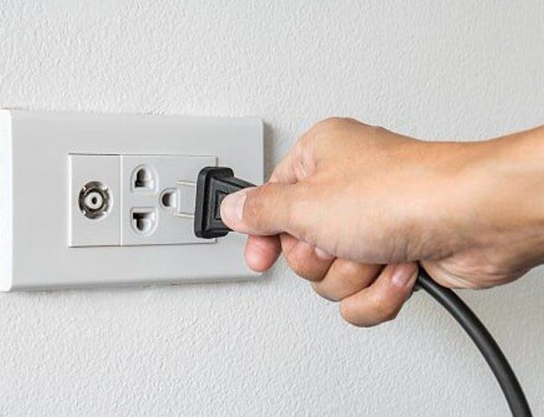
Generally, there are 2 types: the child who bites into the cord producing severe lip, face and tongue injuries and the child or adult who becomes grounded while touching an appliance or other object that is energized.
The latter type is declining in frequency in North America due to the use of ground fault circuit interrupters (GFCIs) in any circuits which supply kitchens, bathrooms or the outside, as these are places where persons may become easily grounded. GFCIs stop current flow if there is a leakage current (ground fault) or more than 0.005 amps (0.6 watts at 120 volts).
- DC:
Direct current injuries are generally encountered when young males inadvertently contact the energized rail of an electrical train system while grounded. This sets up a circuit which produces myonecrosis and electrical burns.
Electrical Injuries
Lab Studies:
- In all patients who by history or physical examination appear to have more than a trivial electrical injury/exposure, obtain the following:
CBC (hemoglobin, hematocrit, white count, red cell indices), electrolytes (sodium, potassium, chloride, carbon dioxide, urea and glucose),creatinine, and urinalysis (specific gravity, pH, color and tests for glucose and hemoglobin). This set gives important baseline values for future treatment.
- In addition to the more common tests, an assessment of muscle damage should be done by ordering:
CPK, total and fractionated, if elevated
Urine myoglobin, if urine gives positive hemoglobin test
Serum myoglobin if the urine is positive for myoglobin
These tests measure the extent of muscle damage in a very effective way. High levels of CPK, identified as muscle with often some elevation in the myocardial component, are seen in any significant exposure to low and high voltage circuits. Lightning rarely will cause an elevation.
If there is extensive muscle damage, there will be myoglobinemia and myoglobinuria.
- In any cases where there is arrest or loss of consciousness, arterial blood gas analysis and a complete drug screen test should strongly be considered.
Imaging Studies:
- Chest X-Ray ( CXR ): If clinically indicated due to chest trauma , shortness of breath or history of CPR at the scene.
- Blunt trauma directly from involuntary contraction of muscles or indirectly from falling secondary to involuntary contraction of muscles, will require imaging studies directed toward discovering possible fractures or even internal injuries.
- These should be approached in the same fashion as with blunt trauma by other causes and appropriate testing should be ordered as indicated.
Other Tests:
- Electrocardiogram (ECG):
An ECG is indicated in any person who is suspected to have electrical injury. If arrhythmias are encountered or if patient had a high voltage injury, monitoring is indicated.
- If no arrhythmias are encountered, further ECG studies are not necessary.
- Electroencephalogram (EEG):
An EEG may be indicated in a person who is unconscious or in arrest.
Whether it will need to be done in the ED depends on a number of institutional factors. It} is not critical to early care decision making.
Procedures:
- An intravenous access should be obtained in all persons who have electrical injury. Consider a central line in those with more than trivial burns and in those who were unconscious or arrested in order to monitor fluid status.
- Fasciotomies of burned extremities may be required in high voltage injuries. Consultation with surgeons with experience in electrical burn injury should be obtained early in the high voltage burned patient, as appropriate early fasciotomy may save a limb
Electrical Injuries
Prehospital Care:
The first thing that must be done is to remove the patient from the circuit. Then, patients who are in arrest require Basic and Advanced Cardiac Life Support regimens. Remember, in electrically induced arrest, there is no underlying disease causing the arrest. Therefore, protracted efforts of resuscitation are met with success more often than usual. Patients who are unconscious but not in arrest, require careful ventilatory observation and assistance, if indicated. Patients with burns above the neck need supplemental oxygen because of the high probability of airway and lung damage.
Secondary blunt trauma is often encountered due to falls caused by involuntary muscular contraction. It is dealt with identically to any other blunt trauma.
Emergency Department Care:
Patients with electrical burns should be stabilized and considered for immediate transfer to the nearest burn center. If such facilities are not available, physicians with experience in burns, preferably in electrical burns, should assume care of the patient.
- All patients with burns and no apparent CNS abnormality should be hydrated. Using the ordinary rule of thumb for treating the ordinary burn patient may result in significant dehydration. In CNS normal patients, administration of physiologic fluids such as Ringer's Lactate at a rates of 10 ml/kg/hour are reasonable during the initial resuscitation.
- In patients with CNS abnormality, hydration must be tempered with the possibility of worsening cerebral edema. There is no easy way to titrate this clinically difficult area.
- Patients who have elevated CPK's and/or myoglobinemia should have mannitol or furosemide added to their regimen to provide diuresis for the toxic myoglobin. This can help to prevent acute tubular necrosis and renal failure secondary to myoglobinuria.
- The lightning strike patient should be treated based on the CNS symptoms. If consciousness is present on admission or returns in the ED, in-patient therapy may not be required. If CNS abnormalities persist, hospitalization is indicated.
- The successfully resuscitated patient exposed to low voltage without significant burns may also be handled primarily on the basis of CNS symptoms and CPK results. If consciousness returns, the CPK is no more than two times normal with negative hemoglobin in the urine and the pulse is regular, hospitalization may be only for brief time periods.
Irregularities of pulse, electrocardiographic changes, myoglobinuria or CNS abnormalities all require hospitalization.Consultations:
Patients with electrical burns require treatment by burn specialists. Prompt transfer to the care of such an individual is indicated. In high voltage electrical burns, early fasciotomy may be indicated to improve circulation. Thus, guidance, as rapidly as possible, should be sought concerning when to initiate this procedure in the emergency department.
- Trauma/Critical Care
- General Surgery
- Plastic/Burn Surgery
Electrical Injuries - MEDICATION
Hydration is the key to reducing the morbidity of electrical injury. If muscle damage is significant, the use of an osmotic diuretic is also indicated.
Drug Category: Fluids
Loss of intravascular volume through the damaged epithelium, as well as loss into extravascular spaces requires fluid resuscitation. This is best be acheived with Lactated ringers.
Drug Name
- Lactated ringers - It is essentially isotonic and has volume restorative properties.
Adult Dose
- Generally administer 10 ml/kg/h during initial resuscitation.
Pediatric Dose
- Use the same regimen as in adults.
Contraindications
- The major complication of isotonic fluid resuscitation is interstitial edema. Edema in an extremity is unsightly, but not a significant complication. Edema in the brain or lungs is potentially fatal. The major contraindication to isotonic fluid resuscitation is pulmonary edema in which the added fluid promotes more edema.
Interactions
- No significant drug interactions have been reported with this product.
Pregnancy
- C - Safety for use during pregnancy has not been established.
Precautions
- Fluid resuscitation will be expected to exacerbate cerebral edema.
- Fluids should be stopped when the desired hemodynamic response
Drug Category: Osmotic Diuretics
If myoglobinemia and myoglobinuria are present, acute renal failure can be minimized by the addition of mannitol to the regimen of fluid resuscitation.
Drug Name
- Mannitol (Osmitrol) - It is an osmotic diuretic which is not significantly metabolized and which passes through the glomerulus without being reabsorbed by the kidney.
Adult Dose
- 50-200 g/24 h IV
- Adjust the dose to maintain a urinary output of 30-50 mL/h
Pediatric Dose
- Under 12 y: Safety and efficacy have not been established.
- However, trial doses of 0.2g/Kg IV followed by careful monitoring of urinary output may be prudent, again with the goal of producing diuresis in the child with myoglobinuria
Contraindications
- Well established anuria due to severe renal disease.
Severe pulmonary edema.
- Active intracranial bleeding except during craniotomy.
Severe dehydration.
- Progressive renal damage or dysfunction after institution of mannitol therapy, including increasing oliguria and azotemia.
- Progressive heart failure occurring after institution of mannitol therapy
Pregnancy:
- C - Safety for use during pregnancy has not been established.
Precautions:
- Severe electrolyte imbalance and dehydration can ensue if a careful monitoring of electrolyte status is not performed.
Electrical Injuries
Further Inpatient Care:
- Inpatient care will be required for burns and for patients with CNS abnormalities. Burns require case specific treatment done by persons with experience and training.
Further Outpatient Care:
- Lightning:
- Patients released from the ED with good CNS function but with otoscopic abnormalities should be referred to a person experienced in treating ear disease and injury. All patients should be referred to an ophthalmologist for evaluation of possible cataract formation, which is reported to occur after lightning strikes.
- Patients without CNS abnormalities, massively elevated CPKs or with electrical burns need no further follow-up. Complete and full recovery is to be expected.
Transfer:
- All patients with history of exposure to high voltage should be transferred for inpatient treatment, preferably by a burn center, on this criterion alone. In addition, mouth burns in a low voltage situation should receive specialized treatment generally available only in burn centers.
- Transfer to an in-patient treatment area should be done if there has not been full return of CNS function, there has been a greater than three-fold elevation in CPK or the presence of myoglobinemia/uria, or there is a persistent arrhythmia.
Deterrence/Prevention:
- Prevention of high voltage electrical injuries requires on-going public education, directed particularly to those in construction trades, using cranes and lifts or exposed to the extreme danger of overhead powerlines. It is particularly important to educate adolescent males regarding the serious nature of electrical distribution equipment.
- Lightning:
- When thunderstorms are in the area, never ever be the tallest object. Avoid golf courses and open fields. Do not stand besides trees. Seek shelter in buildings or cars. If caught outdoors, lie on the ground.
Low voltage:
- Never ever use appliances which give you a shock, until they are repaired. Encourage the use of GFCI's on all outlets where a person may be grounded but always in bathrooms, kitchens and outside. If using equipment with no built in GFCI, use a GFCI extension cord.
- If there are not significant burns, and if consciousness returns before arriving to or in the ED, full recovery is usual. Rarely, persistent arrhythmias have been recorded. Persistence of unconsciousness leads to a graver prognosis. Full recovery is not expected if unconsciousness persists for 24 hours.
Complications:
- Lightning:
If consciousness is regained before arriving, or inside the ED, a full recovery is expected. Prolonged unconsciousness leads to a graver prognosis. Full recovery is not expected if unconsciousness persists for 24 hours.
Low Voltage Mouth Burns:
- With proper treatment, the disfigurement of low voltage mouth injuries can be minimized. Scarring will always be present but not extremely disfiguring.
High Voltage:
- Survival with massive burns is now the exception rather than the rule. The incidence of extremity loss has been reduced with improved treatment but has not been eliminated. Severe disfigurement is the rule, even when extremities are preserved due to the massive irreparable destruction of nerve and muscle.
Prognosis:
- For those without burns, prognosis is based upon CNS function. If it promptly returns, prognosis is excellent, even in patients who arrest.
- For those with burns, survival continues to improve with the improvement of burn care. Disfigurement continues to be a major problem.
Patient Education:
- If the cause of the injury is established, obviously counseling concerning avoiding such hazards is important. Generally, the injury speaks more eloquently than we do.
Electrical Injuries : Miscellaneous
Medical/Legal Pitfalls:
Litigation over the injury is to be expected. It is extremely helpful if you document the presence and absence of electrical burns. Diagramming these injuries is always indicated. Photographing the injured and uninjured areas of the body is extremely helpful. It is always proper to have written consent for photographs.
Generally in electrical injuries, there is a solvent defendant other than the medical practitioner. Thus, suits against practitioners in such cases are rare. Documenting the extent of the injuries is, however, extremely helpful should the practitioner end up being the only defendant.
Car Accidents
Neck Injuries
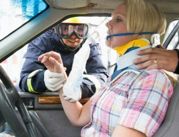
External forces imposed upon the neck can vary from an automobile accident, an athletic injury, a fall, to a direct blow. The extent of injury cannot always be immediately determined and the primary care of the injured patient muse be scrupulously observed.
FIGURE 42. Muscular deceleration and acceleration of the forward flexing r, pine from the erect ligamentous support to the fully flexed ligamentous restriction. Muscle eccentric and concentric contraction permits forward flexion and re-extension. At the scene of an accident it muse be assumed that the neck has been injured and possibly the cord has also been traumatized. Roenegenograms, which are mandatory in severe injuries, should be taken early.
An adequate airway muse be maintained while the patient is still at the place of injury.
Further care of the cervical fracture-dislocation with or without necrologic deficit is beyond the scope of this text and requires the intercedence of a specialist. The precautions listed here are to insure the patient's reaching this specialist without additional damage or injury.
In injuries with no severe impact or symptoms and no necrologic deficit the condition of acute sprain with soft tissue injury muse be assumed to have occurred. An accident need not be severe to cause cervical injury; in face, injury can be sustained from rapid braking of a car, stepping from an unnoticed curb, or stepping into a depression in the ground.
Forceful flexion and/or extension causes damage to the longitudinal ligaments, the intervertebral disk, the areicular capsules, the ligaments, and the muscles of the neck. Simultaneously the spinal canal is acutely narrowed as are the intervertebral foramina. The sensitive tissues of the functional unit are involved, which results in local and referred pain.
Diagnosis and Evaluation - X-Rays and Physical Examination
FIGURE 97. Head and cervical flexion. In head flexion the head is flexed upon the cervical spine with movement only at the occipito-atlas; in neck flexion there is reversal of the cervical lordosis. Most flexion occurs between C4 and C5 or C5 and C6.
Lateral flexion and rotation occur simultaneously when done actively by the patient.
FIGURE 100. Neck flexor trauma from rear-end injury. Hyperextension causes an overstretch and inappropriate contraction of the neck flexors with residual flexor disability.
Treatment of Acute Injury
Because an acute injury may cause soft tissue inflammation with undoubted microscopic tissue damage, these tissues muse be placed at rest in a physiologic position. A muscle spasm follows an acute injury to protect the injured pare by immobilization, but this spasm, as well as being beneficial, may also be detrimental. Therefore, proper immobilization of the neck is mandatory but presents problems.
Immobilization of the neck requires prevention of motion of the occipito-cervical junction and of the cervical column from the second to seventh vertebra. Rotation and lateral flexion muse also be restricted. Immobilization with a cervical collar is indicated.
Collar and Brace
Plastic collars are made more Hoff felt collars by including metal strips that adjust the height of the collar. It is estimated this collar restricts flexion and extension by 75 percent but restricts rotation by only 50 percent.
FIGURE 101. Felt collar recommended for occipito-cervical restriction.
The four-poster cervical brace is cumbersome to maintain and difficult to adjust. It restricts approximately the same degree of flexion-extension as does the plastic collar but permits rotation of the atlaneoaxial joint.
No collar or brace has proven to fully immobilize the neck in all directions. Only a Halo body jacket brace will fully immobilize the neck in flexion-extension, rotation, and lateral bending, and this is regarded only in severe orthopedic or neurologic problems.
Exercises
Exercises should be considered very early in the care of hyperextension injuries-of-significance intensity of the cervical spine.
Physical Therapy
Modalities such as heat, ice, ultrasound, and infrared have their advocates, but usually ice early and heat in later stages of recovery are preferred. Massage of the painful, Bender, and endurated muscles is of value preceding gentle gradual exercises.
Traction
Traction also has equivocal support but has clinical value when properly applied.
Traction with neck in slight flexion will decrease the lordosis and open the intervertebral foramina and separate the posterior articulations.
Traction can also be applied manually which can be combined with lateral and rotatory screech to increase range of motion.
FIGURE 101. Felt collar recommended for occipito-cervical restriction.
FIGURE 104. Ineffective home door cervical traction. The patient is too close to the door to get the corred neck flexion angle.
The door freely opens and closes, not permitting constant traction. The patient cannot extend the legs or assume a comfortable position. This type of home traction is not recommended.
FIGURE 105. Recommended home traction from chinning bar in sitting position.
After subsidence of acute symptomatology by the use of a collar, concomitant exercises and daily traction posture and modification of daily activities must be undertaken. The collar should never be used for prolonged periods nor be used without appropriate exercises.
Burns:
Tissue injury caused by thermal, radiation, chemical, or electrical contact resulting in protein denaturation, burn wound edema, and loss of intravascular fluid volume due to increased vascular permeability.
Sports Injuries
ROTATOR CUFF TEAR
A complete tear may clinically resemble an acute peritendinitis in that there is pain, marked limitation, and tenderness. The arm cannot be abducted at the glenohumeral joint and the patient shrugs.
A partial tear reacts exactly as does peritendinitis with the torn fibers contracting and forming a swelling of the cuff, which obstructs free motion in the suprahumeral space.
Physical Medicine and Rehabilitation Measures
Surgical repair should be considered in a complete tear in a reasonably young person whose activities and profession require full range of shoulder motion with good strength. However, in elderly or severely debilitated patients, surgical repair may not be successful or lasting. With full understanding by the patient of the possible outcome of surgery, every patient, nevertheless, can be considered a surgical candidate. Postoperative care will require a full exercise program as outlined for the other shoulder conditions.
TENNIS ELBOW
Pain and tenderness over the lateral epicondylar region in using the forearm in motion of wrist extension and supination is commonly termed tennis elbow or lateral epicondylitis.
TREATMENT
Wrist immobilization with a cock-up splint or with a plaster cast relieves the tension on the wrist extensors. No immobilization of the elbow is indicated.
The site of tenderness can be injected with a mixture of an anesthetic agent and steroid. The exact site of pathology should be injected.
When all conservative means fail, surgical intervention may be requested.
Sports - Knee Injury
PAIN SYNDROMES
Complaints of pain in the knee must be clarified by clinical manifestations.
Collateral Ligament Pain Syndromes
- A similar force or stress that causes meniscal tears can cause ligamentous tears.
- Ligamentous injuries may be accompanied by meniscus injuries and this fact must always be kept in mind.
- Injury to the cruciate ligaments causes anteroposterior instability.
Ligamentous Injuries
- Ligamentous strains or minor tears actually heal well in time, and active exercises must be begun early.
Meniscal Injuries
- Meniscal tears in the inner two thirds of the meniscus (the avascular zone) do not heal whereas those whose tears are in the outer vascular zone and are reduced can heal.
After successful reduction and adequate immobilization, restoration exercises should be undertaken. The major exercise program should be to strengthen the quadriceps mechanism.
- If conservative physical medicine and rehabilitation measures fail, then surgical intervention is indicated.
- And physical medicine and rehabilitation measures are initiated post operatively for early rehabilitation.


